Fibroblast growth factor
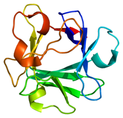
Fibroblast growth factors ( FGF Abbr, ENGL. Fibroblast Growth Factor ) are a group of growth factors, referred to as the FGF family. A total of 23 members of the FGF group known to date: FGF- 1 to FGF- 23rd
FGFs are single- chain polypeptides with a mass of mostly 16 to 22 kDa. They are among the signaling proteins, which are important and potent regulators of cell growth and differentiation of cells. They play a key role in embryonic development. Accordingly, disorders of FGF- functions lead to severe developmental disorders in the embryonic period. In the adult organism, FGFs control gewebsreparative processes, and are actively involved in the processes of wound healing and the formation of new blood vessels (angiogenesis), as well as in the regeneration of nerves and cartilage. FGFs have been detected in almost all tissues of the organism.
FGFs control and change (typically stimulate ) proliferation (multiplication), migration ( migration) and differentiation of cells, particularly endothelial cells, but also from muscle cells ( particularly smooth muscle cells) and fibroblasts. Of the complex process of angiogenesis is controlled substantially by growth factors of the FGF family and influenced. Prototypes of the FGF family is FGF-1 (acidic FGF ) and FGF -2 (basic FGF ).
Mechanism of Action
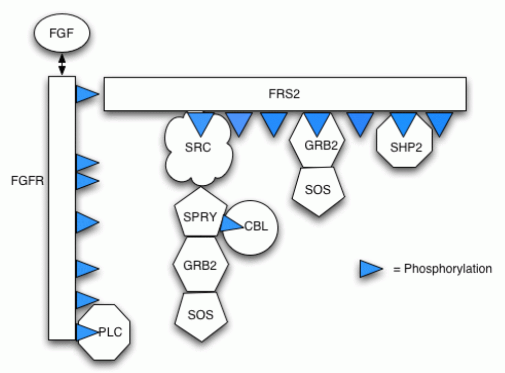
FGF molecules bind to their cognate receptors ( FGFR = FGF receptor) on the cell surface. FGFRs are receptor tyrosine kinases that - are activated by autophosphorylation and intracellular signaling cascade a set with subsequent gene activation in transition - after binding of the ligand FGF. FGFRs consist of an extracellular region that possess three immunoglobulin -like (IG -like) protein Domänes (D1 -D3), a singular transmembrane helix, and an intracellular protein domain with tyrosine kinase activity .. There are four FGFRs: FGFR1, FGFR2, FGFR3, FGFR4. By alternative mRNA splicing of the receptors FGFR1 three additional forms of FGFRs result (a total of seven known FGFRs ), which are denoted by "b" and "c". FGF-1 is the only ligand, which binds to the seven cell surface receptors. The resulting upon binding of FGF and FGFR actual signaling complex at the cell membrane is called the ternary complex, consisting of two identical FGF ligands FGF two identical receptor units and either one or two of the heparan sulfate chains ..
A special feature of the mechanism of action "of FGF is that it ( glycosaminoglycan ) is substantially enhanced by the very high affinity of the FGF for proteoglycans, heparan sulfates and heparin. Therefore, the growth factors of the FGF family were formerly referred to as heparin -binding growth factors ( HBGFs ).
FGFs are secreted physiologically with any form of tissue damage, but especially during hypoxia and ischemia ( up -regulation ), active.
FGF family
FGF-1 (a- FGF) is the most active growth factor of the FGF family. It consists of 141 amino acids. The FGF-1 -encoding gene is located on chromosome 5. Through its extensive binding capacity with all FGF receptors, the biological, cell mitogenic effects are particularly pronounced and characterized by the initiation of cell proliferation, migration and differentiation. FGF- 1 acts on endothelial cells particularly, but also to many other cell types. Due to the particularly pronounced angiogenic activity of FGF -1 FGF-1 recently intensively studied in clinical research, and used in various clinical trials in human medicine .. The plasma half-life of FGF -1 after intramyocardial injection is from 0.4 to 4.6 hours.
FGF -2 (b- FGF) has a similar Molar mass such as FGF -1; the structure determined to be more than 50% in accordance with the FGF -1. The gene encoding FGF-2 is located on chromosome 4. The effects of FGF-2 are similar to those of the FGF- 1, but not quite as pronounced intensive. It is made among others by adipocytes and influences bone metabolism.
FGF-3 consists of 240 amino acids, its structure is about 40% homologous to the FGF -1; the encoding gene is located on chromosome 11. The physiological effects of FGF -3 are still little known, but possibly FGF -3 is especially important during the embryonic period.
FGF -4 ( K -FGF or earlier HST1 ) is composed of 206 amino acids that is 40% homologous to the structure of FGF -1-3, and the coding gene is located on chromosome 11, FGF-4 is commonly found in tumors, particularly in gastric tumors. In normal adult tissues, FGF-4 occurs only in low concentration.
FGF- 5 consists of 251 amino acids, the coding gene is located on chromosome 4. FGF -5 apparently plays during embryonic development is a major ( and Others angiogenic ) role in adult tissues is FGF -5 but only in very low concentrations.
FGF -6 (formerly HST2 ) is 70% homologous to the FGF- fourth The encoding gene is located on chromosome 12. Little is known about its effects, FGF -6 may play a role in wound healing.
FGF -7 was referred to first as a keratinocyte growth factor (KGF ); it has a specific proliferative effect on epithelial cells. The encoding gene is located on chromosome 15
FGF -8 ( Genlokalisation on chromosome 10) may play a key role in the formation of the extremities during the embryonic period.
FGF -9, first referred to as glioma -derived growth factor ( GDGF ), stimulates in particular the proliferation and activation of glial cells in the brain.
FGF- 10 to FGF -22: Although the amino acid sequences and structures of these growth factors are described, little is known about the detailed functions of these proteins. FGF -18 stimulates in model organisms for intra-articular injection into the formation of cartilage. A recombinantly produced human FGF- 18 is in clinical trials.
FGF-23 is secreted by osteoblasts and is a major Regulatur of phosphate and vitamin D homeostasis. FGF-23 stimulates excretion of phosphate through the kidneys. Task of FGF -23 is to keep the phosphate levels in the blood despite varying phosphate intake in the diet constant. Elevated blood levels of FGF -23 lead to a drop in the levels of phosphate in the blood ( hypophosphatemia ), decreased production of 1,25 ( OH) 2- vitamin D and rickets or osteomalacia ( osteomalacia ). Decreased blood levels of FGF -23 lead to increased phosphate levels in the blood ( hyperphosphatemia ), increased production of 1,25 ( OH) 2- vitamin D, soft tissue calcifications, exuberant bone formation ( hyperostosis ) and reduced life expectancy. In renal patients, which must begin with the dialysis treatment, elevated FGF -23 levels are associated with increased mortality (mortality).
History
The first FGFs have been discovered in the 1970s, and their chemical structures described. First, it was assumed that they act solely on fibroblasts ( hence the name). Later, however, it was found that FGFs much more general functions - particularly proliferation and differentiation - have, and can act on almost all cells. Today, even the FGFs are known to have no effect on fibroblasts, such as FGF -7 and FGF- 9th FGF-1 and FGF -2 were first recovered from the brain of bovine, and isolated and later also the structures of human growth factors FGF-1 and FGF -2 have been described.
Early the reinforcing effect of heparin and heparan sulfate has been detected to the function of FGF. To date (2007), 23 different sub-types of the FGF family are described.
Functions and Medical Importance
The various FGF- types have intense mitogenic activity and are of great importance for organ differentiation and development in the embryonic period. They regulate cell proliferation, migration and differentiation. A regular cell and tissue differentiation is not possible without FGF. In adult tissues and organs have FGFs - especially FGF-1 - very intensive activity for the induction of angiogenesis. This property of FGFs has aroused more recently the interest of medical research, since angiogenesis may come as a therapeutic principle in such disease states and disorders to the application, in which a disturbance of the arterial blood flow (atherosclerosis ) is present, such as coronary heart disease (CHD ) and peripheral arterial disease (PAD ). Hypoxia and ischemia triggers the secretion of FGF-1 and FGF- 2, so that there is an up- regulation of FGF receptors in the tissue. Caused by the binding of FGF and FGFR induction of angiogenesis can also be understood in the sense of a repair process, which improves circulation improving. Clinical studies with patients suffering from severe coronary artery disease could not demonstrate FGF-1 induced new vessels in the human heart muscle, as well as a local increase in blood flow with reduction of angina symptoms. Also in disorders of wound healing, such as diabetic ulcers, FGFs act, particularly FGF-1, promoting wound healing. Animal experiments could even be detected a the extent of stroke significantly reducing effect of FGF -1.
Example of FGF-1 -induced angiogenesis in human heart muscle. Left angiographic view of the vascular network in the area of the newly formed front wall of the left ventricle. Right, gray value analysis to quantify the angiogenic effect.
Stress SPECT analysis of the human myocardium by intramyocardial FGF-1 application. Left, before FGF-1 treatment. Law, three months after treatment.
Due to the broad mitogenic activity of FGF they are in current clinical research and investigated with respect to their positive effect on osteoporosis ( osteoblast activation by FGF -1) and with respect to their potential for repair of cartilage damage ( osteoarthritis ). Also, FGF -1 and ( lower) and FGF -2 may be a stimulation of so-called cardiac progenitor cells (cardiac progenitor cells) achieve in terms of maturity of these local, present in the myocardium, the heart muscle precursor cells into adult cardiomyocytes. Finally, a further clinical potential of FGF -1 is its capability of regenerating nerve cells.
Since FGFs are also found in elevated concentrations in many tumors, where they are involved in tumor angiogenesis, to research on the anti- angiogenic tumor therapy depend on an inhibition of angiogenic activity of FGFs.


