Spinal canal
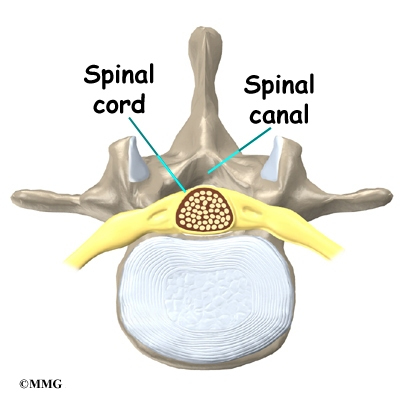
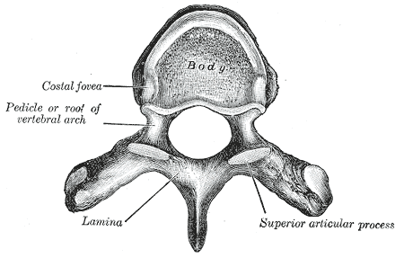
The spinal canal ( vertebral canal ) or spinal canal, also called the spinal canal, the foramen by the superposed ( foramina Vertebralia ) of the vortex formed channel within the spine, where the spinal cord is, and runs from the first cervical vertebra through the cervical, thoracic and lumbar spine to the sacrum. Windwärts belly (ventral ) of the channel alternately through the vertebral body ( corpora vertebrae) and the intervertebral discs ( intervertebral disks ), sideways and move upward (dorsal ) through the vertebral arches ( arcus vertebrae) limited. Between two adjacent vertebrae is found in each case to two sides for the paired segmental spinal nerves, an opening of the spinal canal as intervertebral foramen ( intervertebral foramen ).
Bands
Between the vertebrae are in the area of the spinal canal two kinds of ribbons,
- The posterior longitudinal ligament (Human ) or dorsal (animals ) on the front and bottom of the spinal canal and
- The Ligmenta flava between the vertebral arches of adjacent vertebrae.
Structures in the vertebral canal
The spinal cord is enclosed as all parts of the central nervous system, through the layers of three meninges. Between the outside around the spinal cord dura mater ( dura mater) and the periosteum (bone skin, at the same time the outer layer of the dura mater) that lines the inner wall of the spinal canal, is one with fat and connective tissue and a venous plexus ( plexus vertebral venous ) filled space, the peri- or epidural space.
In the epidural space are surrounded by a ballooning of the spinal dura mater, nerve roots of the spinal nerves and the outgoing dorsal root ganglion. Can these nerve roots off ( epidural ) About an injection of a locally acting anesthetic ( local anesthetic ) in this space.
Also located in this chamber the blood vessels supplying the spinal cord. It takes place on the spinal cord branches ( Rami spinal ) of the vertebral artery, the posterior intercostal arteries ( dorsal in animals ) and the lumbar arteries. These Spina blasphemy pull it over the intervertebral foramen ( intervertebral foramen ) from both sides into the spinal canal and form on the front ( in animals underside ) of the spinal cord an unpaired, extending in the longitudinal direction of the artery, the anterior spinal artery ( in animals as arteria spinalis ventralis called ). It can be regarded as Längsanastomose of segmental spinal cord branches, thus connecting all inflows in the longitudinal direction with each other.
In addition, the corresponding epidural veins form a dense network ( plexus ) of vessels, so the vertebral plexus internus ventralis on the front ( in animals bottom ). This vascular network is particularly vulnerable during surgical procedures near the spinal canal. Bleeding from this plexus often can not be completely silent, which leads to scars later ( arachnoiditis adhaesiva ). Together with the adipose tissue is the epidural venous plexus a cushion for the support of the spinal cord.
Inside the bag- shaped sheath through the dura mater, the spinal cord is surrounded by the two soft spinal meninges ( arachnoid and pia mater ) and the between these broader liquorführenden subarachnoid space. In particular, the two strands of connective tissue per formed laterally to the pia mater anchor as ligament denticulatum the leptomeningeal enveloped spinal cord with pink tapering like trains on the inner surface of the dura.
Diseases
A violation of the integrity of the spinal canal and thus the spinal cord, primarily by a spinal fracture or a herniated disc, often has serious consequences, it may result in paraplegia may occur.
By degenerative changes in the spine the spinal canal can be narrowed (spinal stenosis) and accordingly cause discomfort.
By tearing of the vessels may lead to a hemorrhage between the meninges, which results in pressure on the spinal cord.
Comments
- Bone
- Spinal column
- Spinal cord


