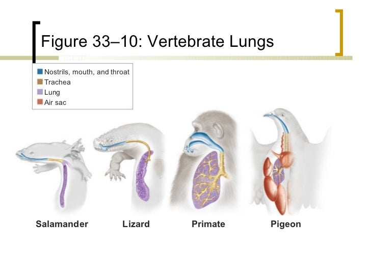
Vertebrate trachea
The trachea and technical in the trachea ( from Ancient Greek τραχύς trachys, rough ', [ note 1 ] is meant " the rough tube ", " the rough artery " - as opposed to the finer blood-carrying vessels) is in vertebrates the connection between the larynx and the bronchial lung. It belongs to the respiratory tract and serves the airline.
Development
In evolutionary terms, the air tube on first in amphibians. Therefore, it can be assumed that this organ was developed about 350 million years ago. The longest trachea there was some dinosaurs around 10 meters in length.
As the entire lower respiratory tract arises the trachea in the embryo of a budding of the foregut.
Anatomy in mammals
The trachea is a flexible tube and begins in the larynx. Here she is at first ventral ( anterior ) of the esophagus and then pulls the neck to her right rib cage. Within the thorax it lies ventral to the esophagus again and divided at the level of 4th to 5th thoracic vertebra in the trachea fork ( tracheal bifurcation ) into the two main bronchi ( bronchi principales ).
In the adult human is 10-12 cm long and 16-20 horseshoe-shaped hyaline cartilage ( tracheal cartilages ), reinforcing its front wall so that it can not collapse even when inhaled. The cartilage rings themselves are by bands - the ligaments anularia - connected to each other, while the rear wall ( paries membranaceus ) connective tissue and muscular ( Trachealmuskel, Musculus trachealis ) structures is made. Through this component, the muscular lumen of the trachea can be narrowed by a quarter.
The perichondrium of the cartilage rings radiates into the ligaments anularia. The mucous membrane of the trachea is firmly adherent to the perichondrium, in the rear wall adjacent part but can move freely, so that may form longitudinal folds in the contraction of Trachealmuskels. Between these folds open glands ( tracheal glands ) forming a seromuköses secretion. About the orderly operation of the pseudostratified ciliated epithelium ( respiratory epithelium ) mucus and dust particles are transported upwards and coughed or abgeräuspert.
In this mucosa lining the entire inner wall of the trachea, also goblet cells and neuro-endocrine cells are distributed, the latter may lie together as neuroepithelial bodies and belong to APUD - system. These are used as those in the bronchi as chemoreceptors for monitoring respiratory gases.
Clinic
The trachea arrived foreign body trigger the cough reflex. Can not be coughed it, the foreign body can lead to suffocation.
An inflammation of the trachea is called tracheitis. You can infectious, allergic or produced by chemical stimuli. Often it is with a laryngeal inflammation ( laryngitis ) combined ( laryngotracheitis ). Softening of the cartilage rings ( Tracheomalacia ) causes the air tube to collapse, especially in the phase of the breathing-in ( see also the dog tracheal ). A narrowing of the trachea can also be caused by space-occupying processes in the area of the trachea and tracheal cancer. Narrowing of the trachea ( tracheal stenosis ) usually lead to a humming noise, which is called stridor trachealis.
The surgical opening of the trachea ( tracheostomy ) is an emergency measure in linings of the upper respiratory tract. In anesthesia to maintain the air flow and the avoidance of inhalation of foreign material into the trachea an endotracheal tube is placed ( endotracheal intubation ).
In birds is the seat of the trachea windpipe worm.


