Bone healing
The bone healing, also called fracture healing after a fracture, depending on the type of fracture ( fracture) and medical care in two ways: as an primary, and secondary bone healing.
History
Albrecht von Haller already recognized in 1767 the importance of the blood vessels of the fracture healing due to an experimental study
Primary bone healing
A primary bone healing can take place only if the broken ends are closely adapted as early as possible after the fracture and they are not mutually movable. This is the case usually only with surgical treatment by internal fixation.
In primary bone healing is no external callus forms. The trabecular bone ( cancellous bone ) grow together by addition of newly formed bone tissue. This bone tissue is formed by activation of osteoblast inner periosteum ( endosteum ). In the area of the medullary cavity of the long bones usually forms an inner bony callus of bone trabeculae. The osteons of compact bone substance ( substantia compacta ) of the two broken ends can and very good adaptation (gap <300 microns ) of two broken ends to grow together and merge again. The neuentstandende bone tissue has initially a lower mechanical strength. It is then reduced from approximately the 8th week by osteoclasts again and aligned by the corresponding pressure and Zugbelastungslinien of the bone ( trajectories) bone tissue replaces (so-called remodeling ).
Secondary bone healing
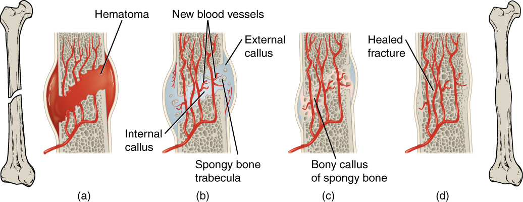
A secondary bone healing occurs in less good adaptation and fixation of the broken ends, as well as with larger bone defects - such as after a comminuted fracture - on.
First exits from the fracture surface of blood and forms a bruise ( hematoma). This leads to activation of the inflammatory cascade and inflammatory cells release cytokines such as interleukin -1 and interleukin-6 released. The blood clot is replaced by granulation tissue and it initially forms a connective tissue scar. First of these processes form a resilient connection of the broken ends and restrict their mobility. By immigrated Knorpelbildner ( chondroblasts ) leads to the formation of fibrous cartilage, which gradually ossified by activated osteoblasts. The resulting sleeve is considerably thicker than the rest of the bone, and is referred to as " callus ". The mechanical strength of callus, however, significantly lower than that of intact bone tissue. Also in the secondary bone healing now uses bone remodeling ( remodeling ), and the callus is gradually reduced and replaced with the corresponding aligned trajectories bone tissue. Depending on the extent of the fracture, the full Knochenausheilung between six and twelve months can take.
If the broken ends do not have enough sedated by the Bindegewebskallus, under no fusion of the broken ends and it may be a false joint ( pseudarthrosis) form. During another break in the immediate vicinity of the first fracture is called a refracture.


