Eyelid
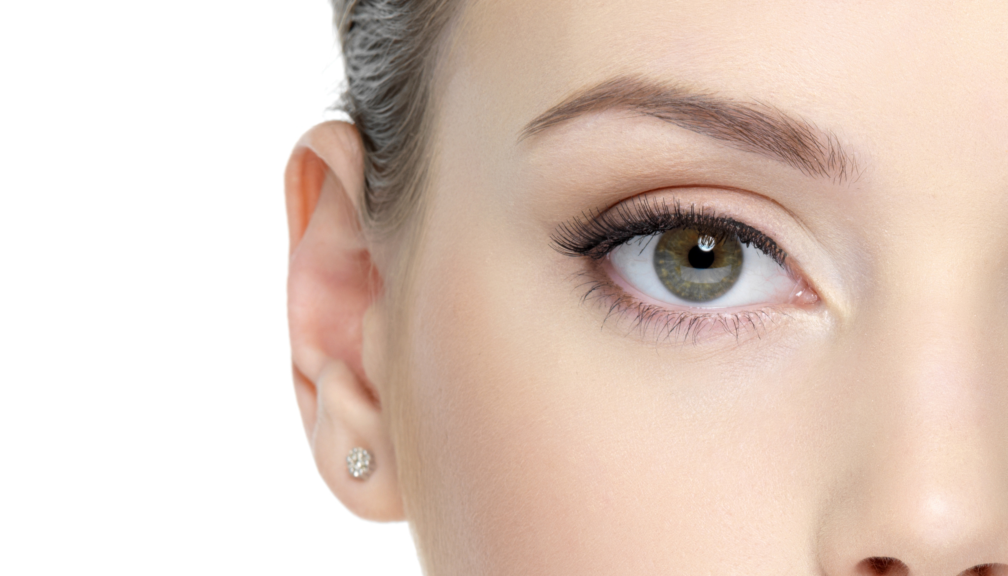
An eyelid (Latin palpebra, Greek Blepharon ) is a thin, made up of muscles, glands, connective tissue and skin existing fold which forms the front boundary of the eye socket (orbit ). There is an upper and a lower eyelid, the opening between the two is called the palpebral fissure. The temples and nose -side location at which both both meet in the open, and when closed is called the angle of the eye, the line between the two Lidwinkeln as Lidachse.
General serve the eyelids protecting and moisturizing the eye. You can cover it completely, thus keep the eye from external influences and his sensitive front sections with the help of tear fluid clean and moist. Composition and structure may differ depending on the species, not all species have eyelids. The depending on the nature of different designs " third eyelid " is called a nictitating membrane.
Some responsible for the movement of the eyelids muscles belong to the group of mimic facial muscles, which underlines the important role of the eyelids in the formation of facial expression and the facial expressions.
- 3.1 Lidstellung
- 3.2 Lidhauterkrankungen
- 3.3 Liddrüsen
- 3.4 Lidranderkrankungen
- 3.5 Movement Disorders
- 3.6 Miscellaneous
- 4.1 Importance of Lidachse
- 4.2 Lidspaltendiagnostik
Function
The eyelids serve two main functions. They are first, to protect the eye from any form of contact, foreign bodies, injuries and light, as well as from mechanical, chemical and otherwise harmful and unwanted effects. The closing and opening of the eyelid is called eyelid, generally also blink. The closing movement is carried out either voluntarily or involuntarily on the blink reflex. This includes the time the relevant stimuli, the eyelids completely ( in humans within an average of about 300 milliseconds ), with its threshold can be reduced by certain diseases. Closing the lid takes place faster than the opening in the rule. On the other hand, a regular blinking ensures that the sensitive cornea (cornea ), which has a substantial proportion of the refraction of light, and thus to an impeccable visual process, as well as the front part of the sclera ( sclera ) at any time sufficiently wetted with tear fluid and becoming dehydrated be preserved. The frequency of the eyeblink in humans is about 10-12 blinks per minute, with women more quickly and more often than men blink.
The closing of the eyelids is synchronous and occurs via the interaction of upper and lower eyelid usually. Exceptions are, for example, hummingbirds and parrots, in which only the upper eyelid is responsible for the closing of the eyelids. In ducks and chickens, however, the closing of the lids birds is performed by a movement of the lower lid to the top.
Additional properties of the eyelids heard that with some exceptions, for example, in rabbits, the eyelids are closed during sleep.
About the protection and supply function, the eyelids play a significant role in the formation of facial expression and the facial expressions. They bear witness to an impression of the current constitution and the total personality deduce characterize it but, for example, a startled or frightened look, as well as the expression of joy, sadness or fatigue. In addition, assume secondary signal functions in some animals nictitating membrane and eyelids.
Anatomy and structure
The eyelids are one of the appendages of the eye. As they consist of a paired organ, often slightly larger, upper eyelid ( palpebra superior) and a lid ( palpebra inferior). Between the two is the palpebral fissure ( rima palpebrarum ). Both eyelids hit the sides together in the canthus ( Angulus oculi or canthus ) and are two bands, the ligamentum palbebrae medial and lateral ligament palbebrae connected to the medial and lateral edge of the eye socket. In the inner canthus there is a nodule- like structure, the Tränenkarunkel.
In the snakes, the locust and some geckos lizard eyelids have grown together and form a corneal surface mounted transparent structure that is referred to as glasses and skinned.
Although cartilaginous fish have eyelids, but they are (eg, nurse sharks ) limited, with few exceptions movable.
Construction
The lid consists of two sheets, an outer and an inner. Said outer sheet lying on the body skin, which is composed of stratified squamous epithelium. Furthermore, the annular orbicularis oculi muscle, and in the upper eyelid levator antagonistic to him, the levator palpebrae superioris, the moving distance is about 20 millimeters in humans, components of the outer sheet. Palpebral Due to its special suspension on the ligament, the inner and outer canthal tendon, the orbicularis oculi muscle does not pull like other ring concentrically, but only causes a closing of the eyelids, without, however, to shorten the palpebral fissure.
The inner layer consists of connective tissue orbital septum, which is located between periorbita palpebrae and the tarsus, and the tarsus itself This is often incorrectly referred to as Lidknorpel, however, is not a cartilage, but one of taut collagen fibers coated by connective tissue. In humans, this has a width of about 25 millimeters, a thickness of about 1 mm and a height of 10 millimeters in the upper eyelid and 5 millimeters in the lower eyelid. This structure is in the upper eyelid, the tendon of the levator palpebrae superioris fan-like at ( Levatoraponeurose ). In the inner sheet more smooth muscle to open and rules of involuntary palpebral fissure is embedded, the musculus tarsalis. He divides in the upper eyelid as superior tarsal muscle, in the lower eyelid as inferior tarsal muscle. On the inner side of the eyelids of the conjunctiva (conjunctiva ) are coated.
From the envelope of the upper lid at the conjunctival fornix, a Oberlidfurche ( palpebral sulcus superior) arise, also known as deck, eyelid or fold. It is formed from the approach of the levator aponeurosis, which starts at the orbicularis oculi muscle under the skin, and is parallel to the lid margin. Their absence indicates an increased subcutaneous fat deposition. A Unterlidfurche ( palpebral sulcus inferior) is often very small, inconspicuous and usually pronounced only when looking down.
In mammals, sitting on the eyelids, the eyelashes (cilia ) in birds bristle feathers ( setae ). They support the protective function of the eyelids, by preventing dust particles and larger foreign bodies from the eye. To the eyelashes, there are several glands:
- Moll glands ( glands ciliares ), named after the German ophthalmologist Jakob Anthony Moll
- Zeis glands ( glands sebaceae ), named after the German surgeon Eduard Zeis
Meibomian and Zeis glands sebaceous glands form as the so-called "Eye Butter ," a secretion that prevents an overflow of tears on the edge of the eyelid. The morning usually dried yellowish secretion residues in the inner canthus be rubbed out of sight in humans colloquially as "sleep". The minor glands produce sweat.
Nictitating membrane
An additional conjunctival fold in the nasal canthus is called a nictitating membrane ( plica semilunaris conjunctivae, membrane nicitans ) or third eyelid ( palpebra tertia ). In humans, it is only rudimentary. In the other mammals it is supported by a cartilage, and so large that they can lie in certain diseases before the entire eye. In many other vertebrates, such as sharks, reptiles and many birds, it is transparent and can be folded as safety glasses to his eyes. In birds, two muscles are in the nictitating membrane embedded, the musculus quadratus membranae nicitantis and the musculus pyramidalis membranae nicitantis. They allow an active blink of the nictitating membrane, which in birds a greater role for the distribution of the tear fluid plays than the actual eyelids. In the domestic fowl the nictitating membrane performs about 35 blinks per minute. The reptiles with fused eyelids missing the nictitating membrane.
Musculature
The following muscles involved in the movement of the eyelids:
Innervation
The sensory innervation is via branches of the trigeminal nerve, the nerve infratrochlearis, supratrochlear nerve and the infraorbital nerve. In addition, the sensitive ulnar nerve supplies the lacrimal the temporal side ( temporal ) canthus. The musculus tarsalis is sympathetic innervation. The outer striated muscle is innervated in the most cases of facial nerve, the nictitating membrane muscles of fine branches of the abducens nerve and the levator palpebrae superioris from the oculomotor nerve.
Blood supply
The eyelids are supplied by the following branches of the ophthalmic artery with blood:
- Supraorbital artery
- Lacrimal artery
- Palpebral artery medial superior
- Artery medial inferior palpebral
The arc- shaped arrangement of the arterial vessels in the upper and lower eyelid is called Arcus Arcus palpebral superior or inferior palpebral. Venous drainage is via the angular vein into the facial vein and the venae palpebral into the superior ophthalmic vein.
Diseases and disorders
Lidstellung
Pathological changes in the Lidstellung found as coloboma, as well as from - or inturned upper or lower eyelid, ectropion and entropion which may be mentioned. The latter can lead to trichiasis, a misalignment of the eyelashes with mechanical irritation of the cornea.
Lidhauterkrankungen
Pathological changes of the eyelid skin may occur as a rash or edema. Pigmentation and fat deposits often exist in the form of xanthelasma. An age-related change in the eyelid skin is known as Blepharochalase. In addition, the eyelids are prone to herpes simplex and herpes zoster infections, as well as other dermatological inflammatory processes.
Liddrüsen
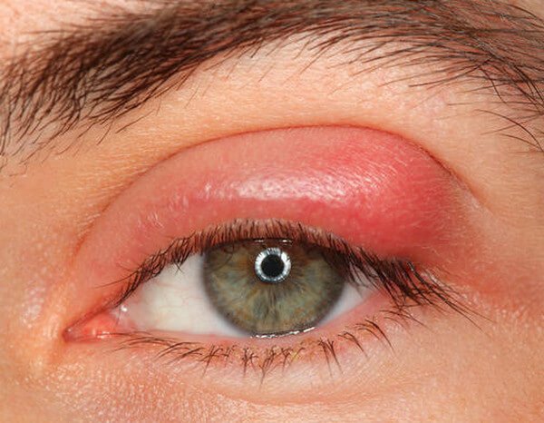
Acute inflammation of the Zeiss or Moll 's glands lead to an external hordeolum ( hordeolum externum ). Are the meibomian glands affected is an internal hordeolum ( hordeolum internum ). Both are summarized under the term of the barley grain. A chronic inflammation of the meibomian glands leading to a chalazion, commonly called hailstone.
Lidranderkrankungen
Typical disease of the lid margin is an inflammation called blepharitis and may have different causes. Blepharitis occurs often in conjunction with a conjunctivitis and blepharoconjunctivitis is then called.
Movement disorders
Movement disorders occur in the form of a partial or complete sale sagging of the upper eyelid (ptosis ) on, coupled with the inability to normally open the eyelid. The congenital or acquired causes may be very different and usually provide a innervationelles problem of innervated by the sympathetic tarsal muscle ( Horner 's syndrome) or dar. by the oculomotor nerve innervated levator palpebrae superioris In contrast to this is referred to by a paralysis of the facial nerve caused inability to fully close the eyelid, as lagophthalmos. This deficit also refers to the blink reflex. A spasmodic closure of the eyelids is called blepharospasm.
Another movement disorder occurs in the form of an involuntary Lidzuckens ( Benign fasciculation ) on, a kind of tremor of one or both eyelids. Most such Lidzucken is indeed annoying, but relatively harmless, even if it lasts for hours or days. It usually disappears on its own. The causes can be varied, ranging from mechanical irritation, fatigue, stress, to mineral deficiencies, especially magnesium deficiency, alcohol consumption or visual stress. Very rarely, a neurological disorder is the cause.
A pathological reduction in eye blink is known as Stellwag character and usually occurs in association with endocrine ophthalmopathy in Graves' disease on. In contrast, an increased blink in the form of frequent blinking is usually the result of mechanical or inflammation-related impairments, such as a foreign body sensation, dry eye, beyond a sign of involuntary muscle contractions ( Tic ) by nervousness.
Another clinical sign of Graves' ophthalmopathy, which manifests itself on the eyelid, is a retired upper eyelid ( retraction ), which gives the impression of a rigid gaze (cooker symbol).
A relative hyperfunction of Lidhebers with striking Oberlidretraktion may result in a paretic limitation of elevation and views of the rectus superior, of a vertical rectus muscle. Here, a stronger impulse to glance phrase is indeed only partially implemented by the affected muscle up approach does not work fully on the synergistic movement of the levator palpebrae superioris out, resulting in an abnormal pulling of the upper lid.
Others
There are a number of tissue changes or tumors known, which can invade the eyelid, including Lidabszesse, cysts, tumors (eg, hemangiomas, carcinoma, basal cell carcinoma or melanoma). At some massive abnormalities of the eyelids can occur when Treacher - Collins syndrome (also: Franceschetti syndrome) have been, a hereditary disease with facial deformities. Other disorders of the eyelids can be caused by parasites. Eyelid edema, thickening of the upper eyelid or Lidhämatome can lead to a pseudoptosis.
One particular sign of Down's syndrome and other genetic disorders is the nasal crease ( epicanthus ), but also physiologically, particularly in Asia, occurs and is therefore again called back and Mongolian fold or double eyelid crease.
Ablepharie denotes the absence of an eyelid. One problem with age yielding of the connective tissue is called Schlupflid and viewed in some cases as cosmetically unfavorable and therefore in need of correction.
The nictitating membrane may also be affected by specific diseases ( see: Diseases of the nictitating membrane ).
Investigation
The inspection of the eyelids is generally carried out with the naked eye, with the help of magnifying glasses or by means of a slit lamp. For the study of the bulbar conjunctiva and eyelid margins often enough pulling apart the upper and lower lid. To assess the palpebral conjunctiva on the bottom beyond the upper eyelid can be up to Tarsus or be folded back up to the upper fold. This is done either by using a simple cotton or glass rod, or with a Desmarres - Lidhalter ( after the French ophthalmologist Louis -Auguste Desmarres ). The method is called a simple and double eversion. In order to keep the lids open in investigations or surgery and preventing unwanted eyelid closure, an eye speculum is used, which keeps open the upper and lower lids, without restricting the access and the top view of the examination or treatment area. In support of this is in some cases, and often in combination with a retrobulbar an artificial paralysis of the orbicularis oculi, a Fazialisblock brought about.
Importance of Lidachse
A special course of Lidachse may be helpful in the diagnosis of disabilities.
A laterally rising Lidachsenverlauf ( inclination of the Lidachse outward above) is often found in people with Down syndrome ( trisomy 21), trisomy 3 and Zellweger syndrome.
A side (lateral ) sloping Lidachsenverlauf ( inclination of the Lidachse outwards below), as represented by hypoplasia ( underdevelopment ) or absence is favored by cheekbones, is common in people with Rubinstein - Taybi syndrome, Pierre Robin syndrome, Noonan encountered syndrome, Marfan syndrome, DiGeorge syndrome, fetal alcohol syndrome, Hallermann - Streiff syndrome, Otopalatodigitales syndrome, Cri syndrome, Cohen syndrome and cat eye syndrome.
Lidspaltendiagnostik
The assessment of the eyelids ( palpebral Rima ) is particularly useful in the clinical picture of Graves' ophthalmopathy for assessment of status and history. Here are the following symptoms:
- By proptosis -related, excessively widened palpebral fissure ( Dalrymple 's sign)
- Lagging of the upper eyelid at infraduction ( von Graefe 's sign)
All these findings can be quantified for a more precise assessment: Dalrymple and von Graefe sign in millimeters, Stellwag characters in blinks per minute.
A clinical sign of myasthenia gravis, the fatigue of the Lidhebers dar. To prove the Simpson - test is performed, in which the patient a longer Aufblick is required, the results of positive results in an increasing droop of the upper lid. After administration of Tensilon these symptoms temporarily disappear again.
Operations on the eyelid
Ophthalmology is holding a series of conservative and surgical procedures for medically indicated local treatment of eyelid diseases, removal of chalazion and tumors, anomalies and movement disorders. Surgical procedures, for example, interventions in the levator palpebrae superioris with ptosis, different techniques of advancement flap, free skin grafts, and upper and Unterlidverlängerung and lateral Lidspaltenverkleinerungen. Another technique for using a special Lidspaltenverengung seam is known as Tarsoraphie. Operations in the area of the lid margins apply here because of the functional and anatomical relationships as particularly challenging.
Increasingly, the plastic surgery seeks to find ways to change the cosmetic and aesthetic appearance, such as blepharoplasty to tighten the lids. Here, the superior palpebral sulcus plays a special role, as it forms, among others, the access point for all of the internal structures of the upper lid. The application of the botulinum toxin nerve agent is also distributed, for example for the removal of harmful folds or for the treatment of blepharospasm. In addition LASER methods are used, for example, in the removal of xanthelasma.
Especially in the field of cosmetic surgery there is therefore a need for more information regarding the expectations of the patient in relation to risks and chances of success.
Etymology and Cultural
The term lid comes from the Old or Middle High German ( HLIT or lit) and means " lid " or " closure ".
Due to an ancient Japanese legend that grew an evergreen tea plant from the two eyelids, the monk Bodhidharma, the Japanese character茶( Cha ), both meaning " tea " has, as well as " eyelid ".
In many cultures it is one of the last services on the people to close the eyelids on the deceased.
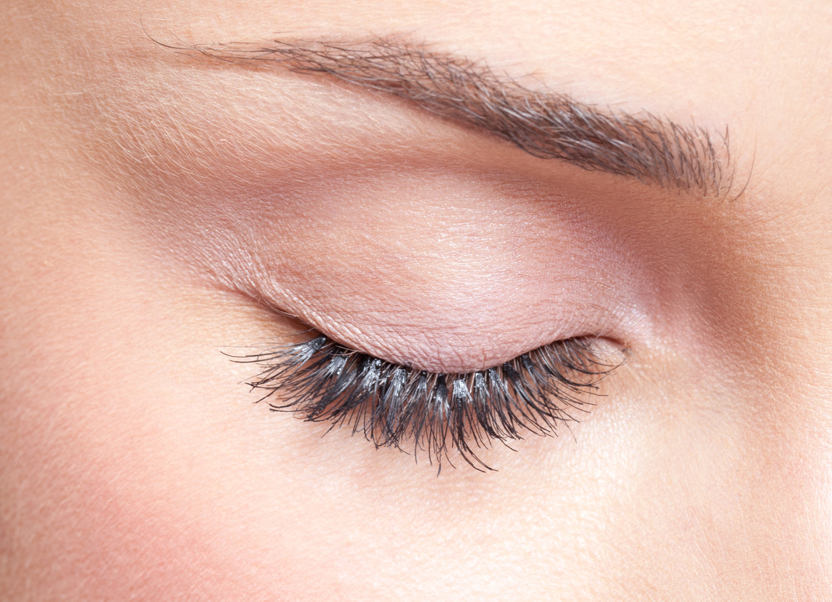
For cosmetic purposes, in particular the upper eyelid is highlighted with eyeliner and / or eyeshadow.


