Taste bud
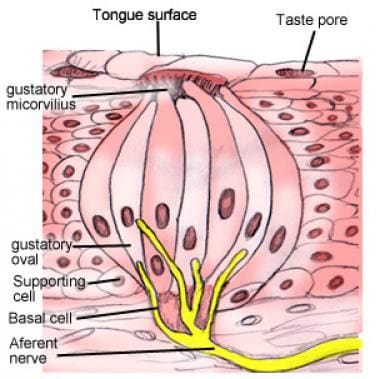
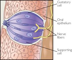
The taste buds or Schmeckknospen ( Caliculi gustatorii ) are onion-shaped structures in the oral mucosa of vertebrates. They accommodate, among other cell types, the sensory cells of taste.
At the top of each taste bud the surrounding epithelium of the mucous membrane forms an opening ( pore gustatory ), can get to the taste sensory cells through the saliva and dissolved therein food items. In these Geschmacksporus superior membrane protrusions of the sensory cells whose apical microvilli, carry the bulk of the molecular taste receptors. At the foot of the entire taste bud the dendrites can be found that afferent neurons, which relay the taste information to the central nervous system. In a taste bud usually contains receptors for several taste qualities (→ Gustatory perception).
In mammals, there are about 75 % of the taste buds in papillae on the tongue, the most on the posterior third of the tongue base to. The rest of the taste buds distributed over the soft palate, nose, pharynx, larynx and upper esophagus. The taste buds of the tongue are assigned to specific surface structures, the taste buds ( papillae gustatoriae ) are called.
To ensure a differentiated taste perception, the buds are cleaned by own Spüldrüsen again, which are named after their discoverer Ebner- Spüldrüsen.
Taste buds
Up to one hundred taste sensory cells lie in a taste bud. More than one hundred taste buds can be in a Geschmackspapille again. The adult man usually has less than one hundred taste buds on the tongue and a total of about almost 10,000 taste buds, most of them in the papillae.
Its form is different to the actual taste buds into three types:
- Papillae ( vallate papillae ): In humans, about a dozen papillae are located in the posterior third of the tongue. They are very much larger than, and may include several hundred Pilzpapillen taste buds.
- Blätterpapillen ( foliate papillae ): The Blätterpapillen in the form of closely consecutive wrinkles. You are on the side of the posterior third of the tongue. Each Blätterpapille contains about 50 taste buds.
- Pilzpapillen ( fungiform papillae ): They are mainly distributed on the anterior two-thirds of the dorsum of the tongue. They carry in humans at about 3-5 taste buds. They are clearly visible when you drank milk.
In addition to these purely mechanical papillae taste buds also come ( papillae mechanicae ) before that receive no taste stimuli. So the Fadenpapillen ( filiform papillae ) are distributed, appealing sensory cells to mechanical stimuli and thus convey a sense of touch on the tongue over the dorsum of the tongue. Very thick and heavily keratinized papillae are conical papillae as ( papillae conicae ), referred to very flat and thick and lenticular papillae ( papillae lentiformes ).
Innervation
Taste receptor cells are secondary sensory cells, they possess a specialized epithelial cells not own axon. To transmit the signals to the central nervous system, they are innervated by afferent nerve, the gustatory fibers. The greater petrosal nerve, a branch of the facial nerve (VII) supplies the taste buds of the palate. Another branch of the facial nerve, the chorda tympani supplies the Pilzpapillen in the anterior two thirds of the tongue and parts of the taste buds in the front Blätterpapillen. The rest of the Blätterpapillen and the papillae are innervated by branches of the glossopharyngeal nerve to the tongue (IX). The taste buds of the epiglottis are supplied by the superior laryngeal nerve, a branch of the vagus nerve (X). Over what nerve are the taste buds of the information of the esophagus and nasopharynx forwarded is not fully understood, it is believed, are that here also the glossopharyngeal and vagus Nervi involved.
Cell types
In every taste bud can be found in humans at about 40-60 sensory cells of taste.
It has long been known that taste buds are formed from a plurality of cell types. The most common division today comprises type I to type III cells and basal cells, which are sometimes referred to as type IV cells. The classification is based initially on observations of tissue sections in the electron microscope and was supported later by molecular biological methods.
- Type I cells are smaller than type II and type III cells, typically electron- dense, have more microvilli on at their peak and have membrane protrusions, envelop the adjacent type II and type III cells. For this reason, a supporting function of these cells is expected. This assumption is supported by the fact that type I cells express, among other things GLAST ( glutamate aspartate transporter ), which is also found in glial cells, the supportive cells of the nervous system.
- Type II cells are less electron dense and have only a single microvillus at its tip to. The precise object of the type II cells is not yet fully understood. Due to the expression pattern is assumed to include a large part of the taste receptors. Type II cells express α - gustducin, among other things, a subunit of the involved in the perception of taste G- protein, phospholipase subtype PLCβ2 and IP3R3, a subtype of Inositoltrisphosphatrezeptors. But even that is important for the transmission of impulses protein synaptobrevin could be detected.
- Type III cells are also less electron dense and form synapses with afferent nerve cells of the three cranial nerves. This is reflected in their protein expression profile: You could synaptobrevin, SNAP -25 - which are involved in transmitter release at the presynaptic terminal - as well as demonstrate NCAM ( neural cell adhesion molecule ). However, there are also important for taste perception proteins PLCβ2 and IP3R3 in type III cells.
- From the basal cells go constantly produces new cells that replace and renew the short-lived sense of taste cells.
Distribution
The distribution and the number of taste buds differ across mammals. In birds the tongue bears no taste buds, here they are localized in the throat. The catfish Ictalurus furcatus even has taste buds all over the body surface.
In 2010 it became known that mammals in the lung appears to have receptors for bitter substances. The inhalation of bitter substances relaxed the smooth muscles of the bronchi should be taught about this, so that the airways are on. These receptors are, however, formed by cells of the airway smooth muscle itself, which can also respond to bitter substances with a dilation; ( integrated neuronal) of sensory cells, one should not speak in this context also.


