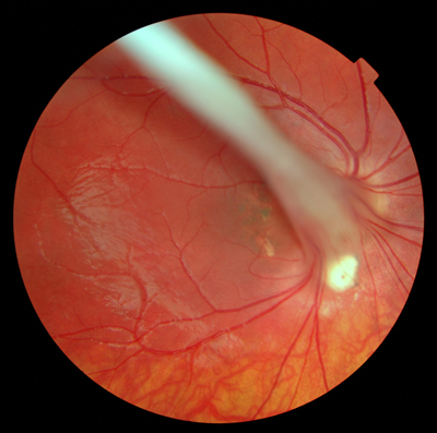
Hyaloid artery
The hyaloid artery or arteriole Hyaloidarterie is one in the eye for the blood supply of lens (9) and vitreous (10). It is a terminal branch of the central artery of the retina pronounced and extends from the exit point of the optic nerve (13 ) to the rear pole of the lens, where it divides into the back of a procedure called the tunica lentis vasculosa vascular network around the lens. In the glass body is surrounded by the trained as Gliahülle Canalis hyaloideus (15 ), which is named after its describer Jules Germain Cloquet Cloquet also channel. The hyaloid artery is present normally only during embryonic development to the nutrient supply to the developing lens and usually disappears shortly before birth.
On the inner side of the lens, after the regression of the hyaloid artery as a designated Mittendorf spot or hyaloid corpuscles turbidity remain, which are generally of no clinical significance. A failing, incomplete or delayed involution is called the hyaloid artery hyaloid artery or persistens persistence and applies in humans and in animals as a rule, harmless abnormality that has little or no visual impairment result. However, they can also lead to restrictions of the visual field and, if it is still bleeding a leader, cause temporary bleeding into the vitreous. Remnants of the tunica lentis vasculosa can cause disturbances in visual acuity on the retina (18). A shrinkage of the canal hyaloideus is a possible reason for a retinal detachment.


.jpg)

